Table of Contents
Pleased to share another scientific paper that I co-authored with colleagues in Pittsburgh USA, supervised by my mentor @kenknischal.In this paper, recently published online in the Cornea journal, we report a case series of Peters Plus syndrome with deep ocular phenotyping using anterior segment optical coherence tomography and ultrasound biomicroscopy.
Peters Plus syndrome (PPS) is a rare, autosomal recessive congenital disorder of glycosylation caused by mutations in the gene B3GLCT. There are only about 100 reported cases of PPS worldwide! PPS is characterised by bilateral anterior segment developmental anomalies, the commonest manifestation of which is congenital corneal opacity (CCO), as well as multiple systemic congenital anomalies and variable developmental delay. Prior to this paper, a detailed description of the ocular findings was lacking in the scientific literature.
https://pubmed.ncbi.nlm.nih.gov/34629439/
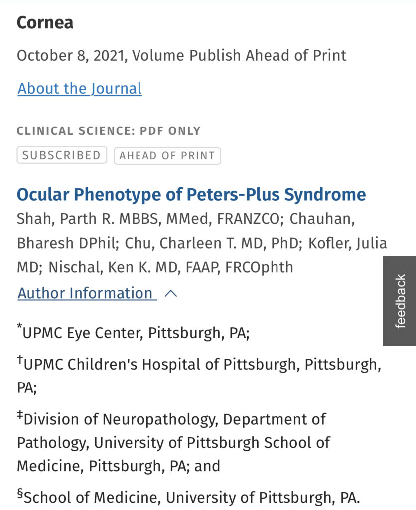
Shah PR, Chauhan B, Chu CT, Kofler J, Nischal KK. Ocular Phenotype of Peters-Plus Syndrome. Cornea. 2021 Oct 8. doi: 10.1097/ICO.0000000000002889. Epub ahead of print. PMID: 34629439.
Abstract
Purpose:
Peters-plus syndrome is a rare, autosomal recessive congenital disorder of glycosylation caused by mutations in the gene B3GLCT. A detailed description of the ocular findings is currently lacking in the scientific literature. We report a case series of Peters-plus syndrome with deep ocular phenotyping using anterior segment optical coherence tomography and ultrasound biomicroscopy. Where available, we describe the histology of host corneal buttons.
Methods:
A retrospective chart review of patients with Peters-plus syndrome was conducted under the care of the senior author between January 2000 and June 2019. Demographic and clinical data including ocular and systemic features, ophthalmic imaging, and molecular diagnostic reports were collected.
Results:
Four cases of Peters-plus syndrome were identified. Three patients were male and 1 was female. Five of the 8 eyes had an avascular paracentral ring opacity with relative central clearing. The paracentral opacity is due to iridocorneal adhesion and the relative central clearing associated with posterior stromal thinning. One eye had persistent fetal vasculature and microphthalmia, which has not previously been reported. One eye from each of 2 patients had a significantly different phenotype with a large vascularized central corneal opacity.
Conclusions:
The most common ocular phenotype seen in Peters-plus syndrome is an avascular paracentral ring opacity with relative central clearing. A different phenotype with a large vascularized corneal opacity may also be observed.