Last Updated: 23 May, 2025
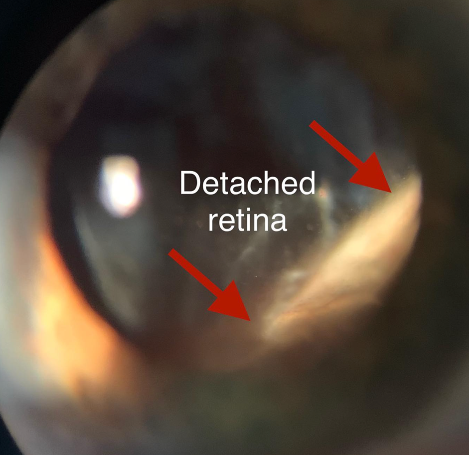
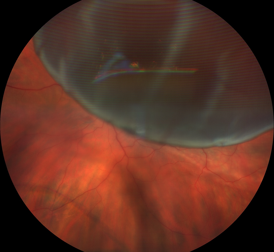
Retinal detachment is an eye emergency in which the retina at the back of the eye pulls away from the tissues around it. It usually occurs due to a tear in the retina, but can also occur in conditions such as diabetic retinopathy in the absence of a tear.
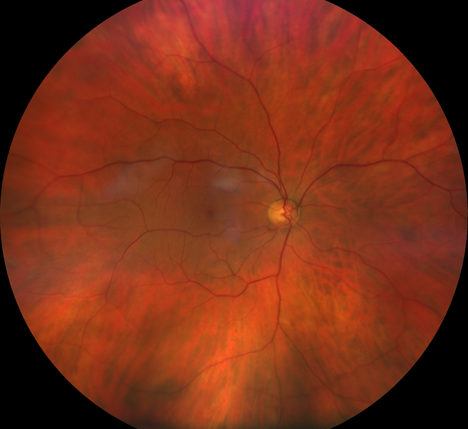
The retina is the light-sensitive film which converts visual input into electrical that are sent to the brain through the optic nerve.
Retinal detachment is a serious condition because it leads to partial or total loss of vision. Treatment as soon as possible is necessary since the layer underneath the retina provides it with nourishment and oxygen. Without urgent treatment, permanent vision loss could occur.
Symptoms of retinal detachment
The symptoms or signs of retinal detachment are:
● A sudden onset of “floaters” in the vision from one eye. Floaters are tiny specks, like flies or insects, that are seen in the visual field.
● Photopsiae, i.e. a sensation of seeing very brief flashes of light, usually in the peripheral vision.
● A curtain-like shadow that starts from one side progresses to cover the visual field.
● Constriction of peripheral vision.
● Blurred vision.
● Loss of central vision.
If you are suffering from any of these symptoms, it is recommended that you see an eye doctor as soon as possible.
Risk of retinal detachment?
You are at higher risk of retinal detachment if you:
- are near-sighted (myopic)
- have had prior eye surgery, such as cataract surgery
- had a retinal tear or detachment in the other eye
- have a family history of retinal detachment
- have had a prior eye injury
- have previous peripheral retinal degeneration or tears
Causes of retinal detachment
Several types of retinal detachments exist.
Rhegmatogenous (Regma-TODGE-Uh-nus).
This is a common type of retinal detachment, which occurs due to retinal hole or tear causing fluid to enter and build up underneath the retina and forcing it out of contact with the underlying tissues. The area of detached retina does not function properly, leading to a loss of visual field. Vitreous separation from the retina, which occurs commonly with ageing, can cause a retinal tear, which can then lead to a rhegmatogenous retinal detachment.
Tractional :
This sort of retinal detachment can arise when tissues there is contraction of tissue on the inner surface of the retina (on the side of the vitreous jelly), causing the retina to pull away. Tractional detachment may occur in patients who have advanced diabetic eye disease.
Exudative :
Fluid collects below the retina, in this kind of detachment. but there are no tears or holes in the retina. Exudative retinal detachment may occur because of trauma, eye tumors or inflammatory conditions affecting the eye.
When to See an Eye Specialist?
Retinal detachment is a medical emergency that requires immediate attention. Recognising the signs early and seeking prompt treatment can significantly impact the outcome and preserve your vision. Here is when you should see an eye specialist:
- Sudden Onset of Significant Floaters: If you suddenly notice a significant increase in floaters—tiny specks or strings that seem to float in your field of vision—this could indicate a retinal tear or detachment.
- Flashes of Light: Experiencing sudden flashes of light, particularly in the peripheral vision, can be a sign that the retina is being disturbed, or is in the process of detaching.
- Curtain-Like Shadow: If you see a curtain-like shadow or a grayish veil that progresses from the periphery toward the center of your vision (from any direction), this is a red flag for possible retinal detachment.
- Peripheral Vision Loss: A sudden loss or constriction of peripheral vision can be an early symptom of retinal detachment.
- Blurred or Loss of Central Vision: Any sudden blurring or loss of central vision, where you cannot see clearly in the middle of your field of vision, should be evaluated urgently.
- Eye Injury: If you have recently experienced trauma or injury to the eye and notice any changes in your vision, seek medical attention to rule out retinal damage.
- Diabetic Patients: For individuals with diabetes, any sudden changes in vision or the appearance of new floaters or flashes should prompt a visit to an eye doctor.
- Family History: If you have a family history of retinal detachment or other eye conditions, it is important to be vigilant about any changes in your vision and consult with your eye specialist.
Remember: Even if you are not experiencing these symptoms but have significant risk factors for retinal detachment, it is wise to schedule a regular eye exam with your eye doctor. Early detection through a comprehensive eye exam can help prevent serious vision loss and address any potential issues before they become severe.
Treatment for retinal detachment
Treatment for retinal detachment depends on a few factors including how much retina is detached and where in the eye the detached is located. The treatment options are laser surgery, cryotheray, or eye surgery to fix any tears or breaks in the retina. The eye surgeon (ophthalmologist) will discuss the kind of surgery they recommend, its risks, and its benefits with you.
Sometimes, the recommened treatment involves using more than one of these treatments at the same time.
1. Laser surgery (photocoagulation)
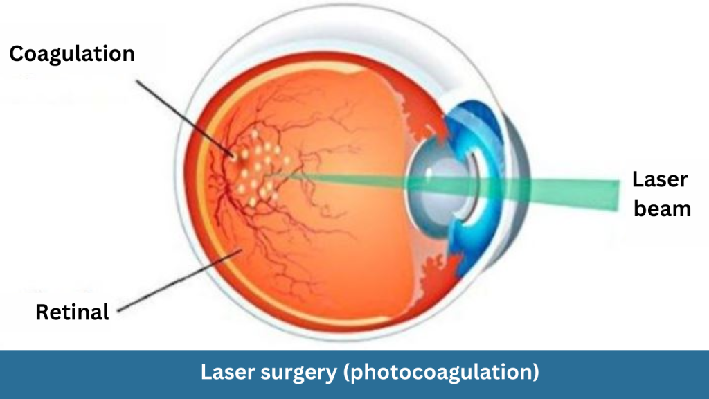
In laser surgery treatment, a medical laser is focused on the retina. This laser makes small burns around the retina hole or tear and small surrounding retinal detachment. Small scars are created because of the burns and this helps to stick the retina down and prevent the detachment from becoming larger.
The procedure involves putting anaesthetic eye drops and focusing the laser through the pupil of the eye. A flashing light may be visible to the patient when the laser energy is delivered.
2. Cryopexy (freezing)
Cryopexy is another way of treating retinal hole or tear. In this procedure, a freezing probe is applied to the outer surface of your eye directly over the tear. Similar to photocoagulation, the freezing causes a scar that helps in holding the retina to the eye wall.
3. Surgery
If your retina is significantly detached, eye surgery in a hospital may be required to situate the retina back to its proper place
For a short time, you may need to stay in the hospital after the surgery. It can be a few weeks’ time before your vision starts to improve after the surgery.
There are 3 main types of surgery to fix a detached retina:
Pneumatic retinopexy:
A small bubble is injected into the eye. The purpose of the bubble is to push your retina back into place so that laser or cryopexy can be effectively applied to repair any holes or tears in the retina. The bubble (if positioned correctly) can push the area of the retina with the retinal hole against the eyewall. This stops more fluid from flowing into the area behind the retina and making the detachment larger. This procedure may be possible in the ophthalmologist clinic. The patient may be required to hold their head in a certain position for several days to keep the bubble’s position in the right place. During a pneumatic retinopexy, the eye is numb using anaesthetic drops. A fine needle is inserted into the vitreous cavity to remove a small amount of fluid, followed by injection of an air bubble. Once the retina is flat, the surgeon will apply laser or freezing treatment to repair holes or tears in the retina. The air bubble will be visible in your peripheral vision after the surgery for some time, and will disappear on its own.
Scleral buckle:
The white of the eye is known as the sclera. During scleral buckle surgery, a tiny, flexible silicone band is inserted onto the sclera. The function of the band is to help your retina reattach by gently intending the sclera toward the detached retina. The buckle is usually left in place but may need to be removed later on. Scleral buckling surgery is performed in a hospital and the eye is anaesthetised fully. The surgeon will also apply laser or freezing treatment to repair holes or tears in the retina.
Vitrectomy:
Vitrectomy surgery involves careful removal of vitreous from the eye. The surgeon will also apply laser or freezing treatment to repair holes or tears in the retina. A gas or replacement fluid like silicone oil may be injected in place of the vitreous. If silicone oil is used, it may need to be removed a few months later.
Recovery from retinal detachment surgery
Immediately following surgery, eye discomfort may persist. Depending upon what happens, the eyelid may feel dry. Eye drops are used to prevent infections. Your vision can get blurred a few days or even weeks after surgery. The ophthalmologist will determine the activities you must avoid immediately after surgery. It may vary by type of surgery. It is likewise determined by your work experience or job type. If a patient has surgery, he/she must tell the ophthalmologist.
Posturing :
It may be important to keep your head in a certain position for a week or two after the surgical operation, if a gas bubble has been injected into your eye. If this is wanted, your ophthalmologist will give an explanation for a way to lie or sit down and for how long. It is critical that you follow their commands to make sure your eye heals nicely.
Sometimes you may need to posture with your head down to ensure that the gas bubble maintains contact with the central retina (macula).
Tips for posturing :
In case you do need to posture, you will generally want to spend 50 minutes out of every hour face down. A day off from posturing is commonly allowed for ingesting, the use of the bathroom and instilling eye drops.
It is generally not necessary to sleep completely straight. Some people can sit in the correct position while sitting on a chair.
– Keep your head down while sitting on the table
– Keep a reading material like a book or an iPad on your lap and read
– lying on your side in bed with pillows propped on either side of you.
Posturing equipment :
You should also not forget to hire a posturing gadget that can support your back, shoulders and neck.
Prevention of retinal detachment
Regular eye checkups can be very helpful when checking your vision. Often trauma occurs when the retinas are detached. The use of protective eyewear for work and play can be helpful in preventing an eye accident. If your vision is affected by flashes or floats, your eye doctor may suggest the procedure. Not everyone with retina needs to have treatment. Treatment of retinal tears or holes may involve laser or cryoscopes.
Long term outlook for retinal detachment :
The success rate of successfully repairing retinal detachments with a single operation is dependent on the type or extent of detachment, and how long it has been there.
Conclusion
Retinal detachment is a vision threatening eye emergency. A comprehensive dilated eye exam by an ophthalmologist can detect retinal tear or localised detachment. If the detachment is caught early, there is a better chance of the treatment options working.
Author Bio
Dr Parth Shah is an ophthalmologist known for his excellence in providing treatment to all eye conditions, including retinal detachment. He is thorough in his knowledge of modern techniques and implements them through individualised treatment for all of his patients.
FAQs