Last Updated: 31 January, 2026
Our eyes are truly remarkable organs, allowing us to perceive the world around us with incredible precision. But what happens when the left and right eyes do not quite work in harmony? That is where eye muscle disorders come into play.
In this article, we will explore some common eye muscle disorders that may necessitate surgical intervention. These conditions, such as simple strabismus, nystagmus, and paralytic strabismus, can significantly impact visual function and eye alignment.
Understanding their specific characteristics and the surgical solutions available is crucial for those affected by these conditions, or those caring for someone who is.
Symptoms of eye muscle disorders
Eye muscle disorders affect the alignment of the eyes and their ability to work together. When the six extraocular muscles that control eye movement become weak, tight, or uncoordinated, the eyes may not move in sync. This leads to a variety of noticeable symptoms depending on severity and cause.
Common symptoms include:
- Double vision (diplopia) caused by the brain receiving two different images from misaligned eyes.
- In children, double vision may go unnoticed because the brain shuts off or suppresses one image, which can lead to lazy eye (amblyopia).
- In children, double vision may go unnoticed because the brain shuts off or suppresses one image, which can lead to lazy eye (amblyopia).
- Blurred vision.
- Difficulty focusing.
- Eye strain, especially during reading or prolonged digital device use.
Additional symptoms may include:
- Headaches
- Dizziness
- Poor depth perception, making activities like driving or climbing stairs more difficult.
- Abnormal head posture (tilting or turning the head) to maintain single vision.
Signs often seen in children:
- Squinting
- Closing one eye
- Turning or tilting the head sideways
Early recognition of these symptoms is key to preventing long-term visual problems.
Causes of eye muscle disorders
Eye muscle disorders can develop from a range of congenital (present at birth) or acquired causes.
Congenital strabismus, present at birth or shortly after, often results from improper development of eye muscles or nerves controlling eye movements. In many cases, the exact cause is unknown, but genetics play a significant role.
Acquired cases may develop due to eye muscle weakness, nerve palsies, or neurological disorders that interfere with muscle coordination.
Medical conditions associated with eye muscle disorders:
- Thyroid eye disease
- Myasthenia gravis
- Diabetes
- Hypertension
- These conditions can affect the nerves supplying the eye muscles, leading to sudden misalignment and double vision
Other contributing factors:
- Eye injuries or trauma.
- Complications from previous eye surgery.
- Uncorrected refractive errors in children, particularly farsightedness.
- Can cause over-focusing and inward turning of the eyes (accommodative esotropia)
- Can cause over-focusing and inward turning of the eyes (accommodative esotropia)
- Viral infections.
- Inflammation
- Stroke, particularly in adults.
Identifying the underlying cause is essential for choosing the most appropriate treatment and reducing the risk of recurrence.
Types of strabismus
Strabismus is one of the most common eye muscle disorders and occurs when the eyes do not align properly. It can be constant or intermittent, and the type varies depending on the direction of eye deviation.
- Esotropia
- One or both eyes turn inward.
- Common in children.
- Often associated with significant farsightedness or congenital muscle imbalance.
- One or both eyes turn inward.
- Exotropia
- One, or both eyes drift outward.
- May be more noticeable during fatigue or illness.
- Intermittent exotropia (eye occasionally turns outward) is especially common in older children
- One, or both eyes drift outward.
- Vertical deviations:
- Hypertropia
- One eye sits higher than the other.
- Often caused by muscle weakness or nerve palsy.
- One eye sits higher than the other.
- Hypotropia
- One eye sits lower than the other.
Vertical deviations frequently cause double vision, and may lead to head tilting to compensate.
Specialised forms of strabismus:
- Paralytic strabismus
- Caused by nerve damage.
- Caused by nerve damage.
- Restrictive strabismus
- Occurs when an eye muscle becomes tight or scarred.
- Commonly seen in thyroid eye disease.
- Occurs when an eye muscle becomes tight or scarred.
Identifying the type of strabismus helps guide treatment options, which may include glasses, eye exercises, or surgery.
Understanding Eye Muscle Disorders
Eye muscle disorders, also known as strabismus or extraocular muscle disorders, refer to conditions where the muscles responsible for eye movement and alignment do not work in sync. The brain, nerves and eye muscles coordinate the movement of both eyes, allowing them to focus on the same object and maintain alignment. When this complex system malfunctions, it can result in various vision problems, including double vision, reduced depth perception, and a visible misalignment of the eyes.
Understanding the basic anatomy of eye muscles is essential. There are six extraocular muscles attached to each eye that control its movements. These muscles work in pairs, with one muscle contracting while the opposing muscle relaxes to move the eye in different directions. The proper functioning of these muscles is crucial for binocular vision, where both eyes work together to provide a single, three-dimensional image of the world.
While some eye muscle disorders may develop due to underlying health conditions or injuries, others can be present from birth. It is important to note that these conditions can affect people of all ages, from infants to adults.
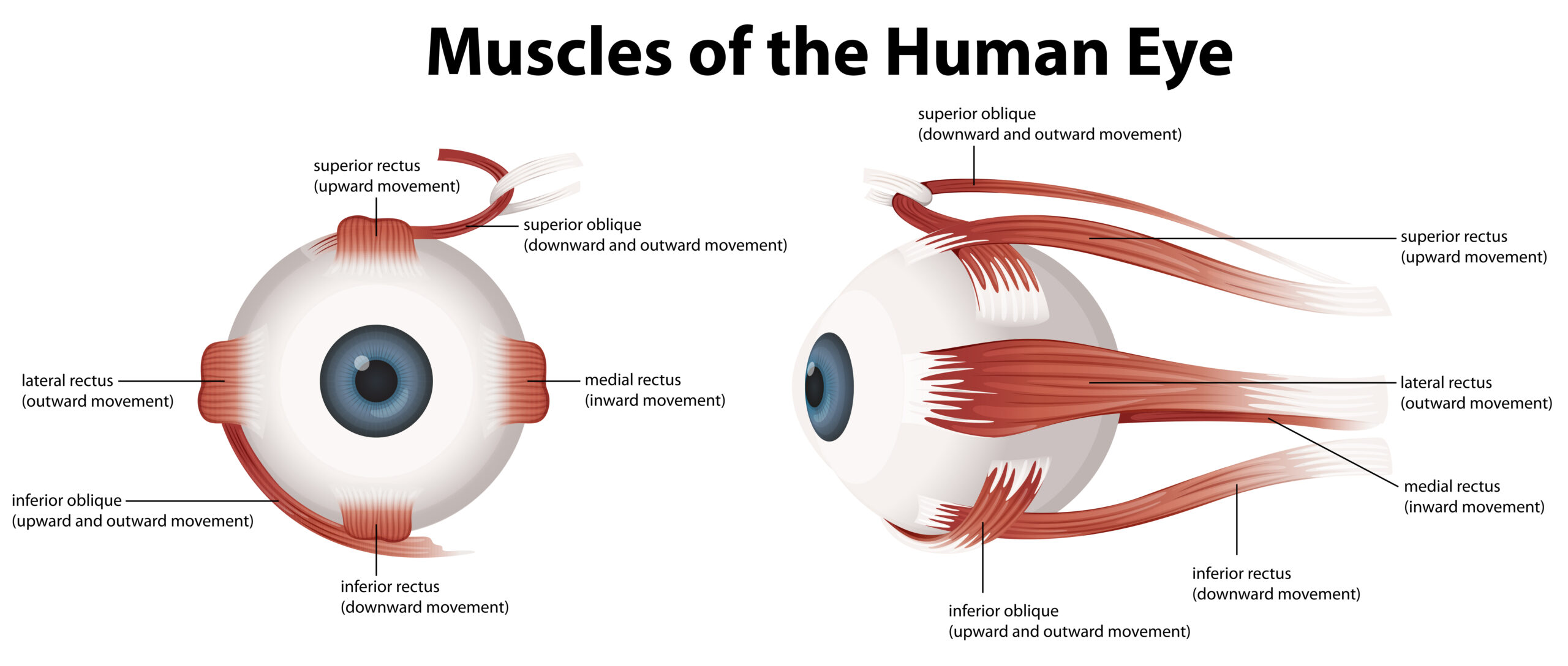
Strabismus – Crossed Eyes
Strabismus, often colloquially referred to as “crossed eyes,” is one of the most common eye muscle disorders. It occurs when the extraocular muscles fail to coordinate correctly, leading to misalignment of the eyes. This misalignment can manifest in different ways:
- Esotropia: In this form of strabismus, one or both eyes turn inward, toward the nose.
- Exotropia: characterised by one or both eyes turning outward, away from the nose.
- Hypertropia: one eye is positioned higher than the other, resulting in vertical misalignment.
Strabismus can have several causes, including problems with the eye muscles, the nerves controlling these muscles, or the brain’s control over eye movements. It can also be related to refractive errors, such as near-sightedness or far-sightedness.
Children and adults with strabismus may experience double vision or amblyopia (lazy eye), which can adversely impact their quality of life.
Nystagmus – Involuntary Eye Movements
Nystagmus is another eye muscle disorder characterised by rapid, involuntary eye movements. These movements can occur in various directions: horizontal (side-to-side), vertical (up-and-down), or even rotational (torsional). Nystagmus can be classified into two main categories:
- Congenital Nystagmus: This type is present at birth or develops during infancy and tends to be stable over time.
- Acquired Nystagmus: Acquired nystagmus develops later in life due to underlying medical conditions, medications, or other factors.
The rapid eye movements associated with nystagmus can make it challenging to maintain steady focus on objects, leading to reduced visual acuity and difficulties with tasks that require visual concentration.
Paralytic Strabismus – Weak Eye Muscles
Paralytic strabismus, also known as paralytic or paretic strabismus, occurs when one or more of the extraocular muscles become weak or paralysed. This weakness can result from damage to the nerves that control the eye muscles or other underlying medical conditions. Paralytic strabismus often leads to limited eye movement and may cause double vision when attempting to look in certain directions.
It is essential to distinguish between paralytic strabismus and non-paralytic strabismus. In non-paralytic strabismus, the eye muscles themselves are typically healthy, but their coordination is impaired. In contrast, paralytic strabismus involves an issue with the muscles themselves, or the nerves controlling them.
Surgical Intervention for Eye Muscle Disorders
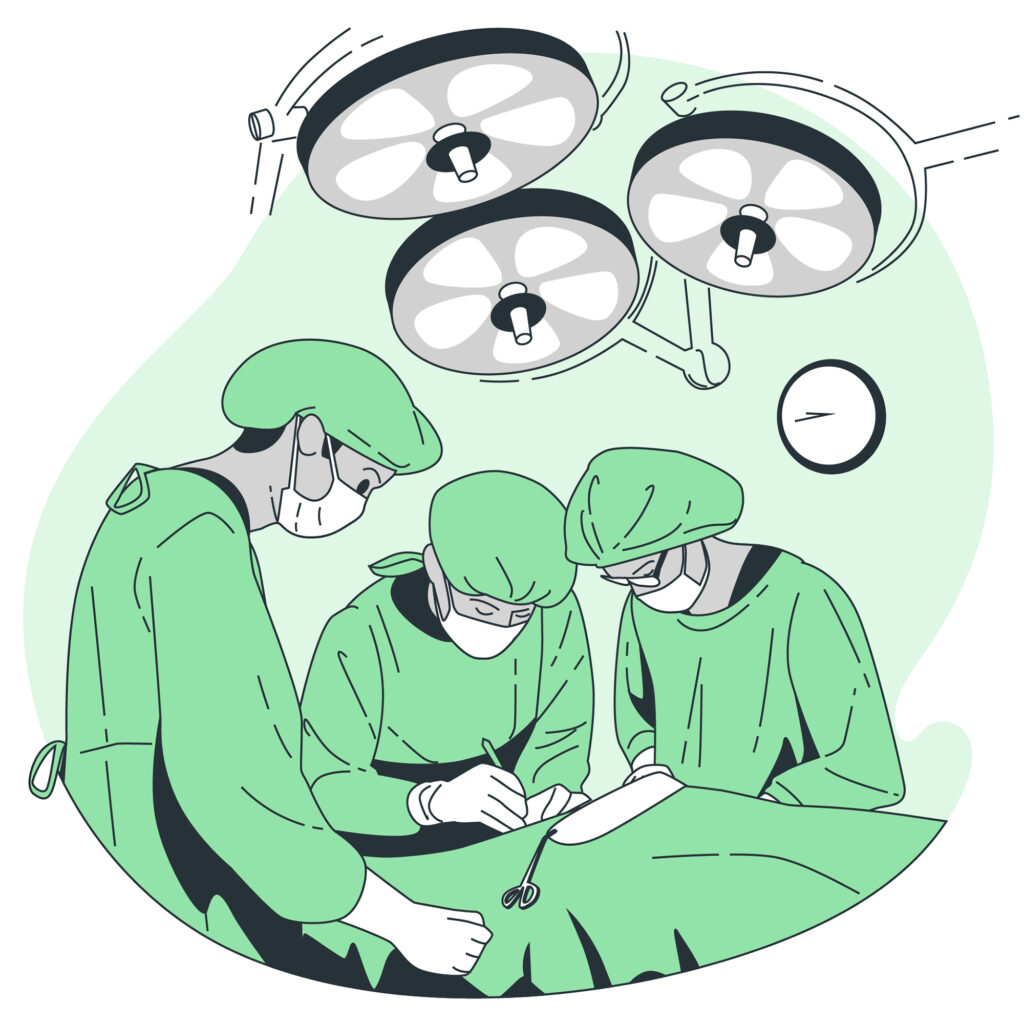
When non-surgical treatments, such as eyeglasses, eye patches, or prisms, do not yield the desired results in correcting eye muscle disorders, surgical intervention may be an option. The primary goal of eye muscle surgery is to restore proper eye alignment and coordination. Different surgical techniques are available, depending on the specific disorder and the individual patient factors.
Here are some common surgical approaches:
- Muscle Recession: In this procedure, an eye surgeon weakens a specific eye muscle by detaching it from the eye and reattaching it farther back. This technique is often used to treat strabismus where an eye turns inward (esotropia).
- Muscle Plication: Plication involves shortening a portion of an eye muscle. This procedure is typically used to strengthen eye muscles.
- Adjustable Sutures: Adjustable sutures allow for precise fine-tuning of eye alignment post-surgery. Eye surgeons can make adjustments after the procedure to achieve the desired eye alignment.
These surgical techniques are performed by ophthalmologists who specialise in eye muscle surgery. The choice of technique depends on the patient’s specific condition and the surgeon’s expertise and experience.
Preparing for Eye Muscle Surgery
Preparation for eye muscle surgery is a crucial step in ensuring the best possible outcome. Patients will typically undergo a thorough preoperative evaluation to assess their eye health, determine the appropriate surgical approach, and address any underlying conditions. The evaluation may include vision tests, eye examinations, and discussions with the surgeon about treatment goals and expectations.
Anaesthesia options will also be discussed with the surgeon. Depending on the patient’s age and medical condition, either general anaesthesia or local anaesthesia with sedation may be recommended for the procedure. The surgeon will explain the chosen anaesthesia method and any associated risks.
Understanding what to expect on the day of surgery is vital for reducing anxiety and ensuring a smooth experience. Patients should receive clear instructions about fasting, medications, and any specific preparations they need to make.
Recovery and Rehabilitation
After eye muscle surgery, the postoperative period is essential for recovery. Eye drops are prescribed for a short duration, and it is important to keep the eyes dry and not to rub the eyes. Recovery timeframes can vary, but patients may experience some discomfort initially. Follow-up appointments are vital to assess the surgical outcome and make any necessary adjustments. In some cases, non-surgical rehabilitation may be recommended to optimise results.
Conclusion
Eye muscle disorders, such as simple strabismus, nystagmus, and paralytic strabismus, can have a profound impact on vision and eye alignment. Surgical intervention is often necessary to address these conditions effectively. By understanding the characteristics of these disorders and the available surgical solutions, individuals can make informed decisions about their eye health. If you or someone you know is experiencing symptoms of an eye muscle disorder, do not hesitate to seek professional advice. Early intervention can lead to better outcomes and an improved quality of life.
Author Bio
Dr Parth Shah is a leading ophthalmologist in Canberra, specialising in eye muscle surgery. With extensive training and experience, he is renowned for his expertise in the field. Dr Shah is dedicated not only to performing successful surgeries but also to patient education. His compassionate approach, combined with technical proficiency, has earned him the trust and gratitude of countless patients. He is a true advocate for eye health and a trusted name in the Canberra ophthalmology community.
FAQs